

Review
History
47 y/o male .
A n intracranial aneurysm was occasionally detected by MR for one month.
Past medical history: No HTN or DM.
Medication: -
NE: (-)
47岁,男性 。
头颅MR检查偶然发现颅内动脉瘤1月。
既往史:否认高血压、糖尿病。
药物:-
神经查体:-

Figure 1. MRI detected a suspicious intracranial aneurysm and no enhancement was observed.
图 1. MRI示可疑颅内动脉瘤,动脉瘤壁未见明显强化。
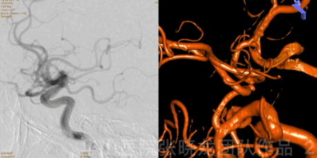
Figure 2 GIF. Compressed left ICA. Right ICA rotational angiogram confirmed an irregular anterior communicating artery aneurysm and the left A1 segment developed well.
图 2 GIF. 左侧颈内动脉压颈。右侧颈内动脉旋转造影证实前交通不规则动脉瘤,同时左侧大脑前A1段发育良好。
1
Strategy
-
An irregular small anterior communicating artery aneurysm with a high rupture risk should be treated.
-
Left A1 segment well developed, therefore the anterior communicating artery can be sacrificed if necessary.
-
-
Solitaire stent will be chosen to straighten the right anterior cerebral artery to reduce recurrence risk.
-
-
前交通动脉不规则小动脉瘤,破裂风险高,建议治疗。
-
-
左侧大脑前A1段发育良好,因此前交通动脉必要时可以闭塞。
-
-
为降低该动脉瘤复发风险,选择Solitaire支架于右侧大脑前动脉释放,拉直右侧大脑前动脉。

Figure 3 GIF. Measurements, An size 2.21*2.29mm, An neck 2.21mm. Right A1 segment proximal diameter 1.89mm, right A2 segment diameter 1.78mm. Headway-21 microcatheter was navigated into right A2 segment. 6F Envoy DA guiding catheter was placed at right ICA petrosal segment. Solitaire 4*20mm stent was deployed from right A2 to A1 segment.
图 3 GIF. 测量,动脉瘤大小2.21*2.29mm,动脉瘤颈2.21mm。右侧A1段近端直径1.89mm,右侧A2段直径1.78mm。Headway-21微导管置于右侧大脑前动脉A2段。6F Envoy DA导引导管置于右侧颈内动脉岩骨段。Solitaire 4*20mm于右侧A2至A1段释放。
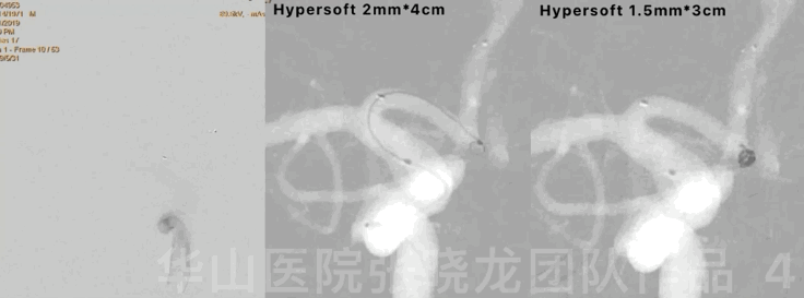
Figure 4 GIF. No bleeding was observed. Then insert 2 coils (Hypersoft 2mm*4cm, Hypersoft 1.5mm*3cm).
图 4 GIF. 复查造影未见出血。然后填入2枚弹簧圈( Hypersoft 2mm*4cm, Hypersoft 1.5mm*3cm )。
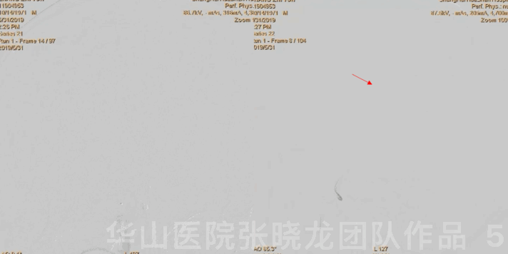
Figure 5 GIF.
The aneurysm was densely packed while a branch of right M3 segment occluded. General heparinization was performed and Tirofiban 14ml was administered.
图 5 GIF. 复查造影前交通动脉瘤致密栓塞,右侧大脑中动脉上干M3段分支闭塞。行全身肝素化,经导引导管给予替罗非班14ml。
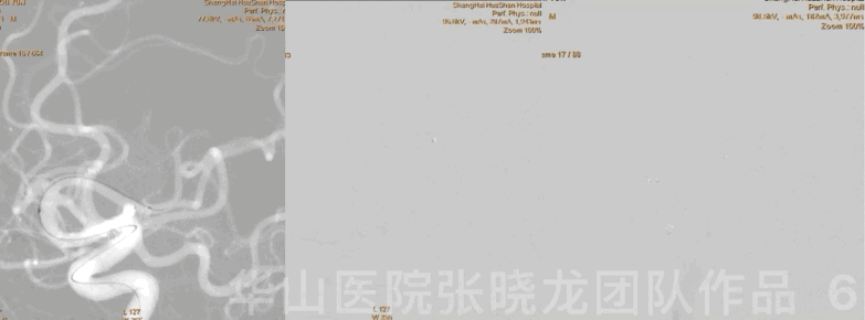
Figure 6
GIF
.
Echelon-10 microcatheter was advanced into the occluded branch and Tirofiban 4ml in total was administered.
图 6 GIF . Echelon-10微导管超选至闭塞分支,经微导管分次给予替罗非班4ml。
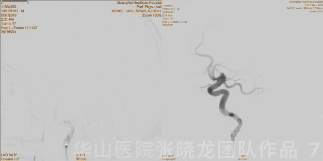
Figure 7 GIF.
Right ICA angiogram showed no relapse of the aneurysm and the occluded branch recanalize partially with pial arteries compensation.
图 7 GIF. 右侧颈内动脉造影显示动脉瘤致密栓塞,闭塞分支血管部分再通,远端软膜代偿良好。
Figure 8 GIF. The aneurysm was not visible and the left anterior cerebral artery was patent from left ICA angiogram.
图 8 GIF. 复查左侧颈内动脉造影动脉瘤未见显影,左侧大脑前动脉通畅。

Figure 9 GIF. Dyna-CT did not detect any hemorrhage.
图 9 GIF. 术后即刻Dyna-CT未见出血。
2
Post Operation
-
NE: GCS 15, no headache, eye movement normal, bilateral muscle strength normal, bilateral Babinski negative.
-
-
Medication:
Tirofiban 13ml/h maintained for 48 hours.
Aspirin 100mg for long term and Clopidogrel 75mg for 3 months.
-
-
查体:神经查体:GCS 15,无头痛,眼球各项运动正常,四肢肌力正常,双侧巴氏征阴性。
-
-
药物:
替罗非班13ml/h维持48小时。
阿司匹林100mg长期口服,氯吡格雷75mg口服3月停药。
-